

Orthopedic Surgery of Cat
Patient presentation and initial physical examination
Ray, a 6 months old male stray kitten was referred to the consultation of Al Barsha Veterinary clinic for injury (fracture) on the left hind limb. The lady who saved him agreed to deposit a financial contribution for the hospitalization and surgery of Ray.
A general physical examination by veterinary for orthopedic surgery showed no major abnormality apart from a non-weight-bearing lameness with the left hind limb. Blood work was found unremarkable.
X-Rays
Two orthogonal views X-ray were performed showing a fracture at the distal tibia (lower extremity of the tibia) of the left hind limb. This type of fracture is common in immature patients and is called Salter-Harris type 1 fracture of physeal fracture.
What is a Physeal Fracture?
It is a fracture located at the level of a growth plate of a long bone (physis) in an immature (growing) animal. The growth plate is made up of cartilage, it is therefore a weak area of the bone that can easily break at that level.
Treatment
If closed immobilization with the application of a cast is possible, it is preferred, however in this case and in this type of fracture, we know they are are unstable by nature and surgical stabilization is preferred and strongly recommended.
Surgical planning was elaborated with several measurements as shown in fig 1.b.

A cross-pinning technique was performed as shown in fig.2. A first pin (Steinmann pin) was introduced from the medial malleolus of the tibia across the fracture line and reaching out to the distal tibia. A second pin was introduced in a diagonal from the lateral malleolus of the fibula across the fracture line and reaching out to the distal tibia.
Post-operative x-rays were done and showed a good reduction and stability of the fracture.
Recovery
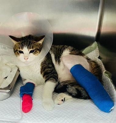
A bandage was applied over the surgical site post-operatively and Ray recovered from anesthesia uneventfully.
Follow-up:
We expect this fracture to heal within 8 to 12 weeks and the two pins will be extracted as soon as the fracture site has consolidated.
Discussion:
In this case, the medial pin (left) is partially within the tibio-crural joint, but we decided to keep it as is to avoid further manipulation and fragilization of the bone fragments. This pin will most likely require to be removed after 8 weeks when the fracture site has completed its healing.
The two pins should have ideally been bent at their lower extremities to avoid possible post-op migration, however, we preferred to not apply any additional bending forces on the bone fragments to preserve them.
Written by: Dr. Mehdi Mzabi, Partner ABVC
Member of the European Society for Veterinary Orthopedics and Traumatology


