
TPLO for Cranial Cruciate Ligament (CCL) in Dogs
Anatomy and function of the cranial cruciate ligament
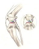
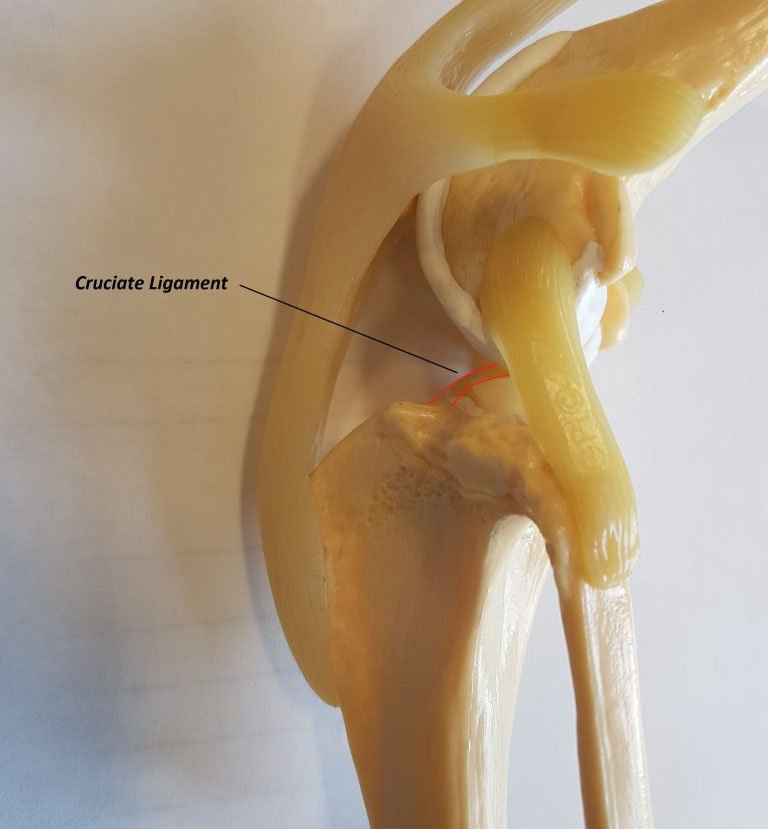
The cranial cruciate ligament (CCL) is located inside the knee joint. It is the same as the human’s “anterior cruciate ligament”. The cranial cruciate ligament runs in diagonal in the middle of the knee and controls back and forth the motion of your cat’s/dog’s knee. The CCL is a ligament of major importance by stabilizing the knee and preventing the tibia from sliding forward in respect of the thigh bone (femur).
 Is cranial cruciate ligament rupture common in dogs?
Yes, CCL rupture is one of the most common causes of lameness in dogs. Unlike humans, the tibial plateau of dogs is extremely steep. In absence of the stabilizing cranial cruciate ligament, the tibia falls completely in front of the femur. The steepness of the tibial plateau puts the CCL under loads of stress during your dog’s walk and run thus explaining the frequency of tibial CCL ruptures in dogs. In dogs, the cruciate ligament tends to undergo degenerative changes weakening the ligament before rupturing completely. This contrasts with people, where a rupture is often associated with an acute injury.
Cranial cruciate disease is seen in all breeds of dogs but certain breeds such as the Labradors and Golden Retrievers, Pugs, Huskies, Maltese, Shih-Tzus, and many others seem to have a higher genetic predisposition. CCL ruptures are less frequent in cats.
What are the signs of a cranial cruciate ligament rupture?
Your dog will exhibit some of the following clinical signs depending on whether the CCL is partially or totally ruptured:
– Lameness
– Pain
– Swelling of the joint
– Stiffness of unwillingness to play and walk
– Reluctance to jump in the car
The resulting instability of the knee damages the cartilage and surrounding bones and leads to a degeneration of the joint’s cartilage called osteoarthritis which is associated with chronic symptoms.
How can your vet confirm a CCL injury?
The diagnosis of a CCL injury is based on a physical and orthopedic examination as well as X-rays of your dog’s knee. Often palpation under sedation or light anesthesia may be necessary to enable the detection of instability of the knee called a “drawer sign” (refer to video with ccl in dog under sedation/anesthesia). Note that a partial rupture of the CCL is a bit more challenging to diagnose. Radiographs provide additional information, especially regarding the presence of osteoarthritis and other changes which may support the diagnosis of a CCL rupture.
Learn more about the TPLO surgery and the aftercare treatment for CCL injury.
Written and updated by:
Dr. Mehdi Mzabi, ABVC Partner and Veterinarian
Member of the French Conseil de l’ordre of Veterinary Doctors, Internship in Small Animal Surgery, and Member of the European Society of Veterinary Orthopaedics and Traumatology


